This work was published in Nucleic Acids Res., Barbe et al., October 28, 2004; 32(19): 5766 - 5779.
Characteristics of the genus Acinetobacter
The genus Acinetobacter is a heterogeneous group which
brings together ubiquitous bacteria found in water, soil, living
organisms and even on human skin. These gamma-proteobacteria are
nowadays classified in the order Pseudomonadales and in the family
Moraxellaceae. Within the genus they are presently divided into 32
genomic species (defined by DNA-DNA hybridization), of which 17 have
been named (situation at the end of 2003). Their taxonomic position
was previously much more confused, as they had been classified in more
than a dozen different genera. The genus Acinetobacter, as to
it, was insufficiently defined for a long time, and has been redefined
(read a short history of the genus
Acinetobacter).
The Gram-negative bacteria which are now classified in the genus
Acinetobacter can be distinguished by the following
characteristics: they are oxidase-negative, catalase-positive,
strictly aerobic and possess a strict respiratory metabolism; they are
immobile with no flagella, do not form spores and appear as cocci
under the microscope (in stationary phase) or as short bacilli, often
in pairs or assembled into longer chains.
Another noteworthy characteristic of numerous strains of the genus
Acinetobacter is that they are capable of utilizing a very diverse range of compounds (1) as sources
of carbon and energy, and of growing on relatively simple media. This
robust metabolism gives them a high capacity for adaptation, which
explains why these bacteria are found in very diverse environments
like their cousins from the genus Pseudomonas. For this reason
they have aroused growing interest because of possible
biotechnological and environmental applications such as bioremediation
(Abd El-Haleem, 2003). Some strains are indeed
capable of degrading or sequestering numerous pollutants, such as
aromatic compounds, hydrocarbons and heavy metals, and of producing
bioemulsifying agents. Other strains of Acinetobacter with a
narrower nutritional spectrum (mainly classified in the species A.
baumannii) have attracted attention because of their
implication in nosocomial infections.
Finally, a specific strain of Acinetobacter has given rise
to considerable interest because of its remarkable competence for
natural transformation: under certain conditions more than one cell in
a hundred may be transformed by DNA from any source present in the
medium, as long as there is a region of homology with this strain’s
genome. This highly transformable strain was obtained in 1969 by
mutagenesis of a strain which had been isolated from soil. First
described under the name BD413, it was renamed ADP1 in 1995 (for
further information, see a short history of the strain
Acinetobacter baylyi ADP1).
Up
Why study the ADP1 strain?
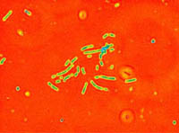 |
Acinetobacter baylyi ADP1 constitutes an interesting model for
the study of competence for natural transformation, which is a genetically
programmed state. Naturally competent bacteria may have a
selective advantage in different ways: supply with nutrients, repair
of mutated alleles by gene conversion, acquisition of new functions by
integration of foreign DNA, ...
When Acinetobacter ADP1 is transformed, double-stranded DNA binds to the cell and is imported as simple-stranded DNA,
even if it comes from organisms which are not related to Acinetobacter (Palmen et al., 1993).
Within the genus Acinetobacter, only strains which can trace their lineage to the soil isolate BD4, such as ADP1, present this natural competence.
The study of the structure and function of the ADP1 natural transformation system have led to the first model of the ADP1 DNA translocator
(Porstendörfer et al., 2000, Friedrich et al., 2001).
Interestingly it comprises several conserved proteins similar to components of type IV pili. Despite these similarities, the two distinct pilus systems of ADP1 are
clearly unlinked to natural transformation. These findings suggest that the ADP1 DNA translocator and type IV pili have evolved from a
common ancestor, but have differentiated into distinct structures with different functions (Averhoff, 2004).
This competence of ADP1 has been exploited in numerous studies. For
example, it is possible to directly transform ADP1 with PCR
products. Nicholas Ornston’s group has introduced DNA fragments from
PCR mutagenesis to study the expression of mutated versions of
heterologous genes (Kok et al.,
1999). Turning to natural diversity, new genes of functional
interest can be recovered in ADP1 using DNA from strains isolated from
soil or other natural environments. Moreover, transformation remains
efficient when ADP1 is placed directly in a sample of humid soil or
waste water (Lorenz et al., 1992),
which makes this strain an excellent model for the study of horizontal
transfer of genes between microorganisms of the
environment. Acinetobacter baylyi ADP1 is even able to develop a
competence state when it colonizes opportunistically a plant infected
by the beta-proteobacterium Ralstonia solanacearum; this has
been exploited to study the possibility of horizontal transfer of DNA
from a transgenic plant to a bacterium (Nielsen
et al., 2000 ; Kay et al.,
2002). It is also envisaged to take advantage of ADP1’s competence
in the soil in order to recover in situ new genes from
non-cultivable bacterial strains. Furthermore, ADP1 remains
transformable when it grows in the form of biofilms. These could
therefore be directly “enhanced” within reactors and would thus
acquire new capacities for biocatalysis, for example in the treatment
of waste water (Hendrickx et al.,
2003).
The condition for natural transformation to
occur remains of course the presence of a sufficient degree of
homology for the exogenous DNA to integrate (Young
et al., 2001). In 1972, Elliot Juni
took advantage of this requirement to validate the boundaries of the
Acinetobacter group; he tested DNAs from 265 strains with very
divergent phenotypes for their capacity to restore growth on minimum
medium of an ADP1 mutant auxotrophic for tryptophan. In this way he
confirmed that all of these strains form a vast genus with ADP1,
excluding oxidase-positive strains.
Finally, the high competence of ADP1 for natural transformation
provides a great opportunity for genetic studies of this
non-pathogenic strain, whose rapid growth on simple media (doubling
times less than one hour at 30o or 37oC) facilitates its
manipulation. It is very easy to transform ADP1 to inactivate a gene,
or to place it under the control of a new promoter. This provides a
convenient method of studying metabolic pathways. These pathways are
extremely diverse in ADP1: like other strains of Acinetobacter
isolated from the environment, it presents a vast nutritional spectrum
(notably, it is one of the rare strains of Acinetobacter which
are able to grow on glucose; see the list of
compounds that ADP1 can utilize as sole carbon and energy source)
and has proven to be capable of degrading numerous compounds. All of
these factors led us to undertake the sequencing of the genome of ADP1
(see Sequencing
project). The annotated sequence has make it possible to perform
comparative genomic studies with related bacteria such as
Escherichia coli and species from the genus
Pseudomonas. Furthermore and above all it has made it possible
to carry out a program of systematic inactivation of ADP1 genes, with
the aim of establishing this strain with its rich genetic repertoire
as a model organism for the study of metabolic transformations in the
environment.
Up
Genomic landscape of Acinetobacter baylyi ADP1
The genome of the Acinetobacter baylyi ADP1 strain is composed of a single circular chromosome composed of 3 598 621 base pairs (bp). Its G+C content (GC%) is 40.3% (the GC% of all the species of the Acinetobacter genus are comprised between 38% and 47%). Surprisingly, this value is quite different from the GC% of the three species of Pseudomonas sequenced to date (GC% from 58.4% to 66.6%), whereas comparisons between 16S rDNA sequences have indicated that Acinetobacter and Pseudomonas are closely related.
The annotation of the genomic sequence of ADP1 was performed on the annotation plaform of the Atelier de Genomique Comparative (AGC) at Genoscope; the data are accessible in the AcinetoScope database. The results of automatic annotation (curves of coding probability) can be visualized using the MaGe graphic annotation interface, together with the choices of the annotator and the synteny data.
The annotation process, which was carried out in two phases (linearly along the chromosome, then by biological function), has yielded 3325 coding sequences (CDS). Their average length is 930 bps; the smallest consists of 69 bps and the longest, of 11 133 bps. We have also identified 76 species of tRNA and 7 rRNA operons, as well as three other RNA motifs, including the RNA components of ribonuclease P and the SRP complex (involved in the signal peptide recognition).
Repeated sequences represent 1.6% of the genomic sequence. Six copies of an insertion sequence of the IS3 family have been identified, in which the GC% does not differ significantly from the overall value for the ADP1 genome. These elements do not seem to have contributed greatly to horizontal transfer events, at least to those in which the donor genomes would have a very different GC%. Genome analysis has also revealed two principal prophage regions. Sixty-four genes have been identified in the longest region (54 kb), of which 45 are unique to Acinetobacter. The others resemble the phage sequences found in Xyllela fastidiosa and Pseudomonas putida. Studies are in progress to determine whether this region still corresponds to a functional prophage. The sequence of the second prophage region, which is 9kb in length, is similar to that of a filamentous phage of Pseudomonas aeroginosa (Pf3).
Up
Orthologs and synteny
Comparisons with sequences in databases have led to assignment of a function to more than 62% of the 3325 protein-coding genes which have been identified in the genomic sequence (56% of these assignments are definitive and 44% are putative). Among the CDS with no assigned function, 675 (20.3% of the total CDS) have been identified as "hypothetical conserved" proteins (homologous to proteins in other organisms with no known function). Finally, 462 CDS (13.9% of the total CDS) are specific to Acinetobacter baylyi ADP1, with no counterpart, and have been designated as "hypothetical". The remaining 3.2% have been annotated as dubious.
As previously hinted by phylogenetic studies based on 16S RNA sequences, the proteomes which are closest to that of Acinetobacter baylyi ADP1 belong to the three sequenced species of Pseudomonas. This result was obtained by searching, in each target genome, the genes with the greatest homology to Acinetobacter CDSs, and by retaining the Acinetobacter CDSs which are their counterpart's most homologous gene in the ADP1 genome (Bidirectional Best Hits, BBH). The ADP1 strain shares a maximum of BBHs (over 63%) with Pseudomonas aeruginosa. This is followed by the two other Pseudomonas species and two other sequenced gamma-proteobacteria, Ralstonia solanacearum and Escherichia coli. Using the percent of CDSs that are syntenic (synteny is here defined as chromosomic colocalization of pairs of orthologous genes in both genomes), P. aeruginosa is again found as the bacterium that is most closely related to ADP1, followed by the previously mentioned species, in the same order. Another indication comes from classification into functional categories: those with the most representatives in ADP1 are enzymatic functions and transport systems. The same clear proximity to ADP1 is again seen with other environmental bacteria, Pseudomonas and the phytopathogenic soil bacterium, Ralstonia solanacearum: these genomes have in common a high proportion of genes associated with inorganic ion transport, with secretion and with biosynthesis of secondary metabolites, which reflects the importance of interactions with the environment. These results are accessible in a table. The results of BBH and synteny studies for 37 bacterial genomes are presented in a histogram.
We have identified an 18 kb region, delimited by very well-conserved synteny groups, which seems to be indicative of horizontal transfer events. The level of conservation is very high with genomes of the phytopathogen P. syringae, with two Vibrio and with the megaplasmid of R. solanacearum. The hypothesis of horizontal transfers between the latter bacterium and Acinetobacter is not new: strains of Acinetobacter from soil, such as ADP1, are adapted to the biochemical diversity created by plants, and are capable of colonizing plant tissues opportunistically during infection by R. solanacearum; in these kinds of niches, transfer of plasmids by conjugation and transfer of chromosomal genes by transformation has already been observed at significant frequencies (Kay et al., 2002).
Up
Functional annotation
More than 60% of the genes of Acinetobacter baylyi ADP1 have been found in synteny groups with the 145 bacterial genomes used for comparison. This conservation has made it possible for us to take into account the gene environment during functional annotation, which proved especially useful in cases in which there was only weak similarity to genes of known function. Some of the most interesting characteristics of the ADP1 gene repertoire (involving several important biological functions) are indicated below:
- Replication: The replication machinery of ADP1 is quite similar to that of E. coli. One interesting difference is the fusion of the dnaQ gene - for which the product in E. coli is responsable for exonuclease 3'->5' activity which corrects errors during replication - and the rnhA gene - which codes for RNase HI, an enzyme which degrades the RNA primers during synthesis of the lagging strand. In proteobacteria, these two genes are often colocalized in a larger synteny group, but fusion of these two genes has never been observed before. It could permit the bacterium to optimize the synthesis of the lagging strand.
- Production of energy: The capacity of ADP1 to grow on glucose, despite the absence of several enzymes of glycolysis (glucokinase, hexokinase and the phosphotransferase system) has already been mentioned. The details of glucose catabolism in ADP1 are presented on another page.
- Aerobic life: As expected, the genome of Acinetobacter baylyi ADP1 which is a strict aerobe, lacks the fnr gene which regulates almost 70 genes implicated in adaptation to an anoxic environment in E. coli. In the same way, it does not have the oxyS gene, whose product is a small antisense RNA implicated in the response to oxidative shock in E. coli.
- Assimilation of nitrites and nitrates: The genes encoding the proteins implicated in the assimilation of nitrates exist as a cluster. One of these genes, for which automatic annotation indicated a similarity with the FAD oxidoreductase of Ralstonia, seems to be a fusion product which encodes the small subunits of nitrite reductase and nitrate reductase. Evidence for this comes from synteny data and the nature of the products of the flanking genes (large subunits of nitrite reductase and nitrate reductase).
- Assimilation of sulfate: The genes which encode the two subunits of ATP sulfurylase are found in ADP1. This enzyme esterifies sulfate with one molecule of ATP to produce adenosine 5' phosphosulfate (APS). On the other hand, APS kinase, which phosphorylates APS to phosphoadenosine 5' phosphosulfate (PAPS) in enterobacteria is absent. The sulfite ion, which is produced in E. coli by reduction of PAPS, seems to be directly produced by reduction of APS in Acinetobacter baylyi ADP1, using an enzyme which is homologous to the PAPS reductase of E. coli, but which carries two motifs which are characteristic of the APS reductases of some plants and bacteria.
- Biosynthesis of polyamines: The absence of polyamines such as putrescine and its derivatives, spermidine and spermine, has already been noted in Acinetobacter. These are replaced by diaminopropane, which these bacteria produce in large quantities. Analysis of the genome has revealed, as expected, the absence of S-adenosylmethionine decarboxylase and spermidine synthetase, two enzymes which intervene in the production of spermidine from putrescine. However, several decarboxylases which should be able to produce putrescine and cadaverine are found in ADP1, whereas these polyamines are barely detectable in Acinetobacter baumannii.
- Polysaccharide metabolism: (results are discussed on another page)
- Catabolic islands: (results are discussed on another page)
- Competency in transformation: (results are discussed on another page)
Up
History of the genus Acinetobacter
The history of the Gram-negative bacteria which make up nowadays
the genus Acinetobacter is complex. These bacteria have been
classified in more than 10 genera, including "Achromobacter”,
Alcaligenes, “Bacterium”, Herellea”, Mima”, Neisseria,
etc. These are ubiquitous bacteria, independently isolated from
different sources by different authors, and for which specific
identification characteristics are lacking. This led to considerable
taxonomic confusion.
The first strains of Acinetobacter were isolated by M.W. Beijerinck in 1911 from soil, and were
named Micrococcus calcoaceticus. These bacteria were
subsequently rediscovered and renamed several times. In 1954 J. Brisou and A.R. Prévot created the genus
Acinetobacter, which brought together, among Gram-negative
saprophytes which did not produce pigments (tribe
Achromobactereae), those that were non-motile. Bacteria which
were obviously unrelated were included in this underdefined genus. In
1957, Brisou designated A. anitratum as the type
species. Application of the oxidase test to bacteria of the genus
Acinetobacter revealed that this genus included both
oxidase-positive and oxidase-negative species. In 1968, P. Baumann and colleagues studied about a
hundred oxidase-negative strains which were in the Moraxella
group (strictly aerobic Gram-negative bacteria with no flagella). They
subjected the bacteria to a numeric taxonomic study in which they
tested growth on 158 different carbon sources. Baumann et al.
demonstrated that these strains were clearly distinguishable from the
oxidase-positive Moraxella, and differed especially in their
much wider nutritional spectrum; they proposed classifying them in the
genus Acinetobacter, redefined as indicated in the
introduction. In 1971, the “subcommittee on Moraxella and
allied bacteria” validated the decision to limit the genus
Acinetobacter to oxidase-negative strains.
Baumann et al. remained prudent regarding the subdivision of
the new Acinetobacter genus. They nevertheless proposed the
existence of three species, including the type species,
A. calcoaceticus. The first DNA-DNA hybridization studies by J. Johnson et al. confirmed both the
clear separation between the Acinetobacter strains and
oxidase-positive strains, and also the heterogeneity of the genus
Acinetobacter. The utilization of the ADP1 strain in transformation tests in 1972 further reinforced
these conclusions. It is still difficult, however, to distinguish
species on the basis of physiological characteristics, and at the
beginning of the 1980s, it was usual to only denote a single species,
A. calcoaceticus.
In 1986, following the first studies of P. Bouvet and P. Grimont, the taxonomy of the
genus Acinetobacter was reorganized by combining the results of
DNA-DNA hybridizations with phenotypic characteristics (for details,
see the exhaustive
review by J.-P. Euzéby (in French)). These numerous studies
finally led to the definition of over 30 genomic species at the end of
2003, of which about half have been named (list of named
species on J.-P. Euzéby’s site). In 1997, the analysis of 16S rRNA
sequences from strains representative of the ensemble of genomic
species described to date confirmed the coherence of the genus and
suggested the existence of five large groups within it (Ibrahim et al., 1997), and in 1999,
phylogenetic studies of the gyrB and rpoD genes
confirmed the relationships of the strains within the genomic species
(Yamamoto et al., 1999).
The genus Acinetobacter was first classified in the
Neisseriaceae family with the genera Neisseria, Moraxella and
Kingella. At the end of the 1980s, DNA-rRNA hybridization
studies underlined the heterogeneity of this family, and J. De Ley
suggested that the Moraxella, Acinetobacter and
Psychrobacter genera should be excluded. In 1991, he grouped
these genera together in a new family, the Moraxellaceae (Rossau et al., 1991).
Up
History of the “ADP1” strain
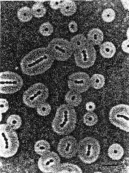 Aspect of Acinetobacter sp. BD4 cells. BD4 is the soil
isolate from which ADP1 was obtained. Wet mount of cells in India ink
reveals the very thick capsule of these bacteria, which appear here as
isolated or chained cocci. ADP1 (BD413) strain is a minicapsulated
mutant of BD4. (photomicrograph from Taylor and
Juni, 1961) |
The origin of the ADP1 strain goes back to the studies of W.H. Taylor and E. Juni in 1960. These two
authors were studying Gram-negative bacteria which were capable of
synthesizing capsular polysaccharides from simple carbon sources. They
isolated a strain of encapsulated Gram-negative cocci from soil which
was capable of utilizing meso-2,3-butanediol as its sole source of
carbon and energy, and called this strain BD4 (for ButaneDiol). The
capsule of BD4 is especially thick (see photo with India ink). When
released into the medium, the capsular polysaccharides, which are
composed of rhamnose, glucose, mannose and glucuronic acid (Kaplan et al., 1985), form a complex with
proteins which acts as an efficient emulsifying agent (Kaplan et al., 1987). A study of sugar
metabolism in BD4, which was initially undertaken to understand the
biosyntheses of these polysaccharides, demonstrated that this strain
can grow on gluconate and glucose despite the absence of the first
enzyme of glycolysis, glucokinase. Glucose-6-phosphate, which is one
of the two first compounds in the biosynthesis of polysaccharides, is
in fact synthesized via the Entner-Douderoff and gluconeogenesis
(Embden-Meyerhoff) pathways, the first step being the oxidation of
glucose to gluconate (Taylor and Juni II and III,
1961). In 1968, when the BD4 strain was classified in the genus
Acinetobacter and in the species A. calcoaceticus, it
came to light that it was one of the rare strains of
Acinetobacter which could use an hexose as its only carbon and
energy source. However, numerous other strains can oxidize glucose or
other sugars (Baumann et al.,
1968).
In 1968, E. Juni and A. Janik wanted to study
further the biosynthesis of the BD4 capsule by isolating intermediary
metabolites in this biosynthetic pathway: after UV- or acridine
orange-induced mutagenesis, they selected several non-encapsulated
mutants which they cultivated in pairs with the goal of demonstrating
complementation by exchange of metabolites. Several of these
co-cultures produced indeed encapsulated cells, but this phenotype
could not be explained by the diffusion of metabolites from one type
of mutant to another: once they were isolated, these cells could
conserve their capsule. Juni and Janik also excluded the hypothesis of
reversion to an encapsulated phenotype. They showed that each
"neocapsulated" mutant had been transformed very efficiently by DNA
from the mutant it was cultivated with, and that the wild-type BD4
strain was also competent for natural transformation (competence was
therefore not related to the absence of a capsule). This fortuitous
discovery appears to be due to the swelling of the unencapsulated
cells cultivated on glucose or gluconate, and to the liberation of DNA
following their lysis. One hypothesis that could explain this swelling
is the increase in osmotic pressure due to the accumulation of
phosphoenopyruvate; this is produced from glyceraldehyde 3-phosphate
which comes from glucose catabolism, and which, in unencapsulated
mutants, would not be consumed in gluconeogenesis.
Juni and Janik noted that two of their mutant strains, BD413 and
BD414, actually had a minuscule capsule. The BD413 strain could be
cultivated in liquid medium without agglutination of the cells, as
seen for cells of other mutant strains or those of wild-type BD4 when
their capsule is removed. The BD413 strain was therefore selected for
further studies on competence for natural transformation because it
was easier to manipulate. In 1985, this strain was renamed ADP1 in the
laboratory of Nicholas
Ornston at Yale University. Its relationship to the species
Acinetobacter calcoaceticus came into question in 1996
following a phylogenetic study based on 16S rDNA sequences (Strätz et al., 1996). As a consequence of
this, it is now designated as Acinetobacter baylyi ADP1.
Up
(1) List of types of compounds which can
be used as sole carbon and energy source to grow certain strains of
Acinetobacter: hydrocarbons, aliphatic alcohols, glycols and
polyols, carbohydrates, non-nitrogenous aliphatic acids and aromatic
compounds, amino acids, amines and various nitrogenous compounds.
(2) The following compounds may be used by
ADP1 as sole carbon and energy source (many other compounds on which
certain strains of Acinetobacter can grow have not been
tested): benzoate, p-hydroxybenzoate, anthranilate, salicylate,
ferulate, vanillate, quinate, shikimate, caffeate, coumarate,
chlorogenate, straight chain dicarboxylic acids, acetate, straight
chain fatty acids, ethanol, lactate, pyruvate, glucose.
Bibliography:
Abd El-Haleem, D. (2003) Acinetobacter : Environmental and Biotechnological Applications. African Journal of Biotechnology 2: 71-74.
Averhoff, B. (2004) DNA transport and natural transformation in mesophilic and thermophilic bacteria. J. Bioenerget. Biomembr. 36: 25-33.
Baumann, P., Doudoroff, M., and Stanier, R.Y. (1968) A Study of the Moraxella Group II. Oxydative-negative Species (Genus Acinetobacter). J. Bacteriol. 95: 1520-1541.
Beijerinck, M.W. (1911) Über Pigmentbildung bei Essigbakterien. Centr. Bakteriol. Parasitenk. Abt. II 29: 169-176.
Bouvet, P.J.M., and Grimont, P.A.D. (1986) Taxonomy of the Genus Acinetobacter with the Recognition of Acinetobacter baumannii sp. nov., Acinetobacter haemolyticus sp. nov., Acinetobacter johnsonii sp. nov., and Acinetobacter junii sp. nov. and Emended Descriptions of Acinetobacter calcoaceticus and Acinetobacter lwoffii. Int. J. Syst. Bacteriol. 36: 228-240.
Brisou, J., and Prévot, A.R. (1954) Etudes de Systématique Bactérienne. X. Révision des espèces réunies dans le genre Achromobacter. Ann. Inst. Pasteur 86: 722-728.
Friedrich, A., Hartsch, T., and Averhoff, B. (2001) Natural transformation in mesophilic and thermophilic bacteria: Identification and charcterization of novel, closely related competence genes in Acinetobacter sp. strain BD413 and Thermus thermophilus HB27. Appl. Environ. Microbiol. 67: 3140-3148.
Hendrickx, L., Hausner, M. and Wuertz, S. (2003) Natural Genetic Transformation in Monoculture Acinetobacter sp. Strain BD413 Biofilms. Appl. Environ. Microbiol. 69: 1721-1727.
Ibrahim, A., Gerner-Smidt, P. and Liesack, W. (1997) Phylogenetic Relationship of the Twenty-one DNA Groups of the Genus Acinetobacter as Revealed by 16S Ribosomal DNA Sequence Analysis. Int. J. Syst. Bacteriol. 47: 837-841.
Johnson, J.L., Anderson, R.S. and Ordal, E.J. (1970) Nucleic Acid Homologies Among Oxidase-negative Moraxella species. J. Bacteriol. 101: 568-573.
Juni, E. (1972) Interspecies Transformation of Acinetobacter: Genetic Evidence for a Ubiquitous Genus. J. Bacteriol. 112: 917-931.
Juni, E., and Janik, A. (1969) Transformation of Acinetobacter calco-aceticus (Bacterium anitratum). J. Bacteriol. 98: 281-288.
Kaplan, N., Rosenberg, E., Jann, B., and Jann, K. (1985) Structural Studies of the Capsular Polysaccharide of Acinetobacter calcoaceticus BD4. Eur. J. Biochem. 152: 453-458.
Kaplan, N., Zosim, Z., and Rosenberg, E. (1987) Reconstitution of emulsifying activity of Acinetobacter calcoaceticus BD4 emulsan by using pure polysaccharide and protein. Appl. Environ. Microbiol. 53: 440-446.
Kay, E., Vogel, T.M., Bertolla, F., Nalin, R., and Simonet, P. (2002) In Situ Transfer of Antibiotic Resistance Genes from Transgenic (Transplastomic) Tobacco Plants to Bacteria. Appl. Environ. Microbiol. 68: 3345-3351.
Kok, R.G., Young, D.M., and Ornston, L.N. (1999) Phenotypic Expression of PCR-Generated Random Mutations in a Pseudomonas putida Gene After Its Introduction into an Acinetobacter Chromosome By Natural Transformation. Appl. Environ. Microbiol. 65: 1675-1680.
Lorenz, M.G., Reipschlager, K., and Wackernagel, W. (1992) Plasmid transformation of naturally competent Acinetobacter calcoaceticus in non-sterile soil extract and groundwater. Arch. Microbiol. 157: 355-360.
Nielsen, K.M., van Elsas, J.D., and Smalla, K. (2000) Transformation of Acinetobacter sp. strain BD413 (pFG4-nptII) with transgenic plant DNA in soil microcosms and effects of kanamycin on selection of transformants. Appl. Environ. Microbiol. 66: 1237-1242.
Palmen, R., Vosman, B., Buijsman, P., Breek, C.K.D., and Hellingwerf, K.J. (1993) Physiological Characterization of Natural Transformation in Acinetobacter calcoaceticus. J. Gen. Microbiol. 139: 295-305.
Porstendörfer, D., Gohl, O., Mayer, F., and Averhoff, B. (2000) ComP, a pilin-like protein essential for natural competence in Acinetobacter sp. BD413: Regulation, modification, and cellular locaalization. J. Bacteriol. 182: 3673-3580.
Rossau, R., Valandschoot, A., Gillis, M., and De Ley, J. (1991) Taxonomy of Moraxellaceae fam. nov., a new bacterial family to accomodate the genera Moraxella, Acinetobacter and Psychrobacter and related organisms. Int. J. Syst. Bacteriol. 41: 310-319.
Strätz, M., Mau, M., and Timmis, K.N. (1996) System to Study Horizontal Gene Exchange Among Microorganisms Without Cultivation of Recipients. Mol. Microbiol. 22: 207-215.
Taylor, W.H., and Juni, E. (1961) Pathways for Biosynthesis of a Bacterial Capsular Polysaccharide I. Characterization of the Organism and Polysaccharide. J. Bacteriol. 81: 688-693.
Taylor, W.H., and Juni, E. (1961) Pathways for Biosynthesis of a Bacterial Capsular Polysaccharide II. Carbohydrate Metabolism and Terminal Oxydation Mechanisms of a Capsule-Producing Coccus. J. Bacteriol. 81: 694-703.
Taylor, W.H., and Juni, E. (1961) Pathways for Biosynthesis of a Bacterial Capsular Polysaccharide III. Syntheses From Radioactive Substrates. J. Biol. Chem. 236: 1231-1234.
Yamamoto, S., Bouvet, P.J., and Harayama, S. (1999) Phylogenetic structures of the genus Acinetobacter based on gyrB sequences: comparison with the grouping by DNA-DNA hybridization. Int. J. Syst. Bacteriol. 49: 87-95.
Young, D.M., and Ornston, L.N. (2001) Functions of the mismatch repair gene mutS from Acinetobacter sp. strain ADP1. J. Bacteriol. 183: 6822-6831.





 home |
overview |
resources |
sequencing |
news |
press releases |
site |
Legal information

