| -> Access to sequencing project |
|
Collaboration
 Annelid Neuroimmunology Laboratory
UMR CNRS 8017
Université des Sciences et Technologies de Lille
Villeneuve d'Ascq, France
|
|
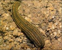
A medicinal leech (photo Annelid Neuroimmunology Laboratory) |
large amount of
research has been conducted on the nervous system of the medicinal
leech, mainly on behavior, synaptic connections and the function of
the ion channels.
These studies have led to a very precise knowledge of the structure of
the nervous system, shown in the diagram below. The leech nervous
system is composed of a chain of ganglia which are linked to each
other by two inter-ganglionic connections composed of nerve
fibers. Thus each segment is innervated by one ganglion.
The nerve chain is ventral and enclosed in a blood sinus. The chain
begins with four ganglia which innervate the anterior portion of the
animal. These ganglia are fused to form two masses. The
supra-esophageal ganglia have a role which is limited to
neurosecretion whereas the sub-esophageal ganglia have an important
sensorial role in innervating the anterior sucker and the three jaws.
These two masses are linked to each other, and form a peri-esophageal
ring. Next, a segmentary ganglion innervates each segment of the
animal's body. Finally, seven fused ganglia form a voluminous caudal
ganglion which principally innervates the posterior sucker of the
leech. The nerve chain is covered by a fibrous capsule which contains
contractile muscle fibers at the level of the connectives.
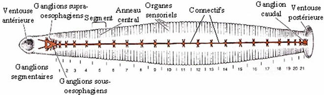
Anatomy of the nerve chain of the medicinal leech (orange).
(Kuffler, S. W. and Nicholls, J. G. (1976) From Neuron to brain. Sinauer Associates, Inc., Sunderland, Massachusetts). |
The neuronal architecture of the segmentary ganglia
has been extensively characterized and is extremely well-conserved
from one ganglion to another. The ganglia contain about 400 cell
bodies each, except for the fifth and sixth ganglia, which are
associated with the reproductive system and which have about
700. Within the ganglia, the cell bodies are separated into 6
follicles, enveloped by a glial cell and numerous microglial
cells. Because of its structural simplicity, the nervous system is a
very accessible tissue, and consequently very useful for in vivo and
in vitro experiments.
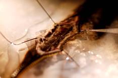
Hirudo
medicinalis's segmentary ganglia (photo Annelid Neuroimmunology Laboratory) |
One of the interesting characteristics of the leech
nervous system is its ability to regenerate following a wound or
lesion.
When a connective is cut, the swimming movements of
the animal are perturbed. Four weeks later, the animal can swim
normally, demonstrating that the regeneration is not only structural
but also functional (1). Numerous in vivo and in vitro experiments
have demonstrated that, unlike mammalian neurons, neurons of the leech
have the ability to regenerate sysapses following a lesion, and above
all, to regain their functions. Recent findings have demonstrated that
microglial cells, as well as components of the extracellular matrix
such as laminin, play an important role in axonal growth and the
formation of synapses during this regeneration. Lesions of the nervous
system also lead to an increase in nitrogen monoxide (NO) and an
accumulation of phagocytic microglial cells at the site of the lesion
(3,4). These experiments suggest that the production of NO may serve
as a signal to inhibit the migration of microglial cells which then
accumulate at the site of the lesion. In fact, the increase in NO is
due to an activation of a NO synthase which is present in the nervous
system after a lesion (5,6). Several experimental observations have
indicated an important role for glial cells of the leech in both
defense and repair of the nervous system.
In the Annelid Neuroimmunology Laboratory, the
research theme devoted to the leech includes four major research axes
which are organized chronologically for the study of the response of
the central nervous system to an experimental lesion [nb: when I did
basic invertebrate biology, we didn't really consider that the leech
had a CENTRALized nervous systen, despite the head ganglia. Maybe this
has changed--S.] :
- Alert, protection and signaling. The factors implicated in
retrograde transport of information.
The innate immune response linked to the lesion.
The signaling molecules which initiate an adaptive response.
- Repair of the nerve chain.
Neurotrophic factors in the leech, their receptors and
partners.
Inhibitors of axonal apoptosis.
- Regeneration of the nerve chain.
Molecules involved in growth and axonal guidance.
- Possible biomedical implications in pathologic or accidental
lesions of the nervous system.
In vitro and in vivo testing of the effects of molecules from the
leech on Invertebrate and higher Vertebrate models.
References :
1. Muller, K. J. and Nicholls, J. G. (1981) Regeneration and
Plasticity. In Neurobiology of the leech (Laboratory, C. S. H., ed.),
pp. 197-226
2. von Bernhardi, R. and Muller, K. J. (1995) Repair of the
central nervous system: lessons from lesions in leeches. J Neurobiol
27, 353-366
3. Chen, A., Kumar, S. M., Sahley, C. L. and Muller, K. J. (2000)
Nitric oxide influences injury-induced microglial migration and
accumulation in the leech CNS. J Neurosci 20, 1036-1043
4. Kumar, S. M., Porterfield, D. M., Muller, K. J., Smith,
P. J. and Sahley, C. L. (2001) Nerve injury induces a rapid efflux of
nitric oxide (NO) detected with a novel NO microsensor. J Neurosci 21,
215-220
5. Salzet, M., Salzet-Raveillon, B., Cocquerelle, C.,
Verger-Bocquet, M., Pryor, S. C., Rialas, C. M., Laurent, V. and
Stefano, G. B. (1997) Leech immunocytes contain proopiomelanocortin:
nitric oxide mediates hemolymph proopiomelanocortin processing. J
Immunol 159, 5400-5411
6. Matias, I., Bisogno, T., Melck, D., Vandenbulcke, F., Verger-Bocquet, M., De Petrocellis, L., Sergheraert, C., Breton, C., Di Marzo, V. and Salzet, M. (2001) Evidence for an endocannabinoid system in the central nervous system of the leech Hirudo medicinalis. Brain Res Mol Brain Res 87, 145-159
|


 home |
overview |
resources |
sequencing |
news |
press releases |
site |
Legal information

